 Fraunhofer-Gesellschaft
High-precision, low-complexity, high-resolution microscopy-based cell sorting
Abstract
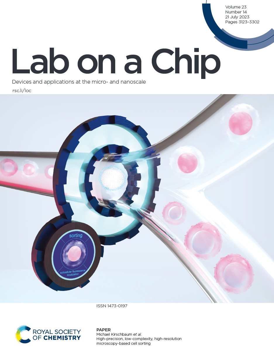
Continuous flow cell sorting based on image analysis is a powerful concept that exploits spatially-resolved features in cells, such as subcellular protein localisation or cell and organelle morphology, to isolate highly specialised cell types that were previously inaccessible to biomedical research, biotechnology, and medicine. Recently, sorting protocols have been proposed that achieve impressive throughput by combining ultra-high flow rates with sophisticated imaging and data processing protocols. However, moderate image quality and high complex experimental setups still prevent the full potential of image-activated cell sorting from being a general-purpose tool.
Here, we present a new low-complexity microfluidic approach based on high numerical aperture wide-field microscopy and precise dielectrophoretic cell handling. It provides high-quality images with unprecedented resolution in image-activated cell sorting (i.e., 216 nm). In addition, it also allows long image processing times of several hundred milliseconds for thorough image analysis, while ensuring reliable and low-loss cell processing. Using our approach, we sorted live T cells based on subcellular localisation of fluorescence signals and demonstrated that purities above 80% are possible while targeting maximum yields and sample volume throughputs in the range of μl min−1. We were able to recover 85% of the target cells analysed. Finally, we ensure and quantify the full vitality of the sorted cells cultivating the cells for a period of time and through colorimetric viability tests.
Gerling, T., Godino, N., Pfisterer, F., Hupf, N., & Kirschbaum, M. (2023). High-precision, low-complexity, high-resolution microscopy-based cell sorting. Lab on a Chip, 2023, 23, 3172 – 3185 https://doi.org/10.1039/D3LC00242J