 Fraunhofer-Gesellschaft
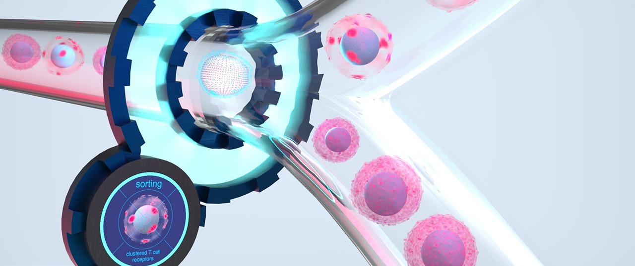
Image-Activated Cell Sorting
Sorting sensitive cell samples based on high-quality microscopy image analysis
Image-activated sorting of small, valuable cell samples overcomes previous limitations of classical sorting methods and enables working at a completely new level of quality

Artistic illustration of image-activated sorting of T cells showing T cell receptor clustering upon activation. Only via spatially-resolved imaging and analysis, the cells with clustered T cell receptors in their membrane can be distinguished from those with homogeneously distributed receptors.
The technological challenge
Separation of heterogeneous cell populations is a key process in modern biomedicine. However, conventional sorting methods based on low-dimensional (»low-content«) sorting criteria such as integral fluorescence intensity or whole-cell scattered light are not able to detect spatially-resolved (»high-content«) features such as the subcellular distribution of proteins, the number and localization of cell organelles or their binding to other cells and use them as separation criteria. To utilize spatially-resolved features for cell sorting, imaging techniques need to be combined with high-precision cell sorting functions. Only then can high-resolution microscopic image data be used together with AI-assisted image analysis to classify and separate cells based on their morphological information.

The table compares various cellular properties with possible use cases that we can address with our approach.
Our approach
In the Fraunhofer-funded IMAGO project, scientists from three different Fraunhofer institutes (Fraunhofer IIS, IOF and IZI-BB) are working together with researchers from Charité Berlin on a microfluidic method in which the microscopic image information of each individual cell flowing through a microchannel is captured and analyzed automatically. According to the separation criterion previously defined by the user, the system generates a sorting decision and precisely separates the target cells from the remaining cells by electrokinetic forces.Our microscope-based approach offers high flexibility in the choice of the imaging technique. Transmitted light, phase contrast or (multi-color) fluorescence imaging with a variety of excitation and emission wavelengths can be easily combined with our approach, as well as less common imaging techniques such as polarization microscopy, quantitative phase imaging and many more. The possibility of image-activated cell sorting in combination with low-loss processing of even small cell numbers overcomes previous limitations of classical sorting methods and opens up new avenues in biomedical research with applications in the fields of immunotherapies and stem cell biology or for the analysis of organoids or small tissue biopsies, and many more.
Fast Facts
- 2-way sorting
- High resolution, high quality imaging data
- 3-4 colors + bright field
- AI-supported cell classification
- »Train by example« using cell images
- Lossless processing of low cell numbers (104 – 106 cells)
- High yield, high purity
- Wide spectrum of cell sizes
- 3…100 µm in diameter
- High biocompatibility
- Low pressure (<4 psi), low shear stress, physiological media, aerosol-free cell deposition
- Single-cell deposition (under development)
- Cell deposition in tubes or plates (or any other vessel)